Mitosis Cell Prophase Metaphase Anaphase Telophase Biology Diagrams Prophase. Most of the time, DNA is tightly coiled and structured around proteins called histones. This packaged form is known as chromatin. The first stage of mitosis sees this chromatin supercoiling from their operational width of 30nm, to the 500nm thickness associated with chromosomes. The material around the nucleoli, contained within the nuclear envelope is DNA in the form of chromatin. This will not pick up a stain well and so will not appear as distinct shapes within the nucleus. Find these indicators of interphase in cell A in the image below. Figure \(\PageIndex{1}\): Cells in an onion root in interphase and prophase.

Interphase The cell spends most of its life in this phase. The DNA in chromosomes copies itself ready for mitosis. Prophase The DNA in chromosomes and their copies condenses to become more visible
Interphase vs. Prophase - What's the Difference? Biology Diagrams
It involves a series of tightly regulated steps, including interphase and prophase. While both interphase and prophase are crucial stages of the cell cycle, they differ in several key attributes. In this article, we will explore and compare the characteristics of interphase and prophase, shedding light on their distinct roles in cell division.
:max_bytes(150000):strip_icc()/interphase-58e3d4a45f9b58ef7e071ea0.jpg)
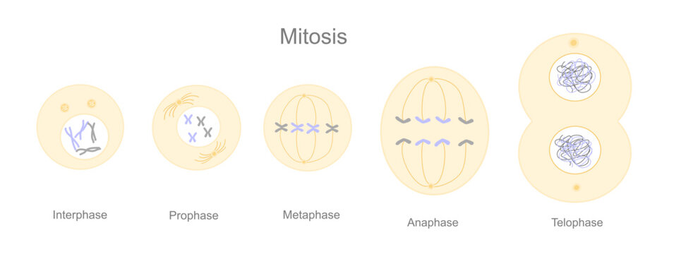
Some cells remain in interphase for days or even years; some cells never leave interphase. At the end of interphase, the cell has duplicated its chromosomes and is ready to move them into separate cells, called daughter cells. This occurs during the four steps of mitosis, called prophase, metaphase, anaphase and telophase. Learn the four stages of mitosis, the process of cell division that produces identical daughter cells. Prophase is when chromatin condenses into chromosomes and the spindle apparatus forms.

The 4 Mitosis Phases: Prophase, Metaphase, Anaphase, Telophase Biology Diagrams
As living organisms grow, their cells must replicate and divide. Most animal cells, except for sex cells, undergo the process of mitosis to create new cells. Through mitosis, one cell creates two genetically identical daughter cells. Mitosis is a complex process consisting of multiple phases; anaphase, interphase, metaphase and prophase. Each phase has its own steps and is integral to the The following points highlight the six main stages of mitosis cell division. The stages are: 1. The Interphase Stage 2. The Early Prophase Stage 3. The Late Prophase Stage 4. The Metaphase Stage 5. The Anaphase Stage 6. The Telophase Stage. 1. The Interphase Stage: The slide shows Interphase stage which is characterized by the following
